The Science of Accessory Tragus: Medical Insights for Riyadh Patients
An accessory tragus, more commonly referred to as a preauricular tag, is a congenital nodule that appears in the area immediately surrounding the external ear. While often viewed simply as a cosmetic irregularity, these structures are rooted in complex embryological development and represent a minor divergence in the formation of the facial and auricular tissues. For many individuals seeking to understand the underlying biology of these growths, Preauricular Tag Removal in Riyadh offers a path toward clinical correction backed by modern surgical and dermatological science. In the Saudi capital, where medical standards align with global benchmarks, patients have access to advanced diagnostic and procedural insights that elevate the treatment from a simple "skin removal" to a precise anatomical restoration. By exploring the medical science behind why these accessory structures exist and how they behave, patients can approach the removal process with a deeper understanding of their own biological history.
The Embryological Origin: A Flaw in Fusion
The development of the human ear is one of the most intricate processes in the first trimester of pregnancy. Between the fifth and ninth weeks of gestation, the external ear (the auricle) begins to form from six distinct mesenchymal proliferations known as the "hillocks of His." These hillocks are derived from the first and second branchial arches. Under normal conditions, these six pieces of tissue migrate and fuse seamlessly to create the complex, curved architecture of the ear, including the tragus, helix, and antihelix.
An accessory tragus forms when there is an "extra" hillock or a failure in the perfect migration of these tissue clusters. Instead of merging into the ear, the tissue remains isolated, developing into a standalone nodule. Because they share the same origin as the main ear, these tags are composed of the same materials: stratified squamous epithelium (skin), subcutaneous fat, and, very often, a core of elastic cartilage. This shared lineage is why they are called "accessory" structures—they are literally "extra" parts of the ear located in an atypical position.
Anatomical Classification: More Than Skin Deep
From a medical perspective, not all accessory tragi are created equal. They are classified based on their composition and their attachment to the underlying facial structures, which dictates the complexity of the removal.
-
Sessile vs. Pedunculated: A pedunculated tag sits on a narrow stalk, making it easier to isolate, whereas a sessile tag has a broad base that is more deeply integrated into the preauricular skin.
-
Cartilaginous vs. Soft Tissue: Many tags contain a central pillar of cartilage. This "root" can sometimes be quite long, extending deep into the subcutaneous fat or even anchoring to the fascia covering the parotid gland.
-
The Preauricular Line: Accessory tragi almost always follow a specific anatomical path known as the preauricular line—an imaginary line stretching from the ear to the corner of the mouth. This reflects the path of the branchial arches during fetal development.
The Clinical Significance of the Location
The preauricular area is a dense anatomical "crossroad." Just beneath the skin where these tags reside lie several critical structures that practitioners must account for:
-
The Facial Nerve: Specifically, the temporal and zygomatic branches of the facial nerve are in the general vicinity. While a tag removal is superficial, a professional clinical approach ensures that the excision depth is precisely controlled to avoid any risk to these motor nerves.
-
The Superficial Temporal Artery: This major vessel provides blood supply to the scalp and face. Its proximity is why professional cauterization and surgical precision are required to prevent hematomas.
-
The Parotid Gland: The largest salivary gland sits just in front of and below the ear. Deeply rooted accessory tragi may sit atop the parotid fascia, requiring a skilled hand to ensure the gland remains undisturbed.
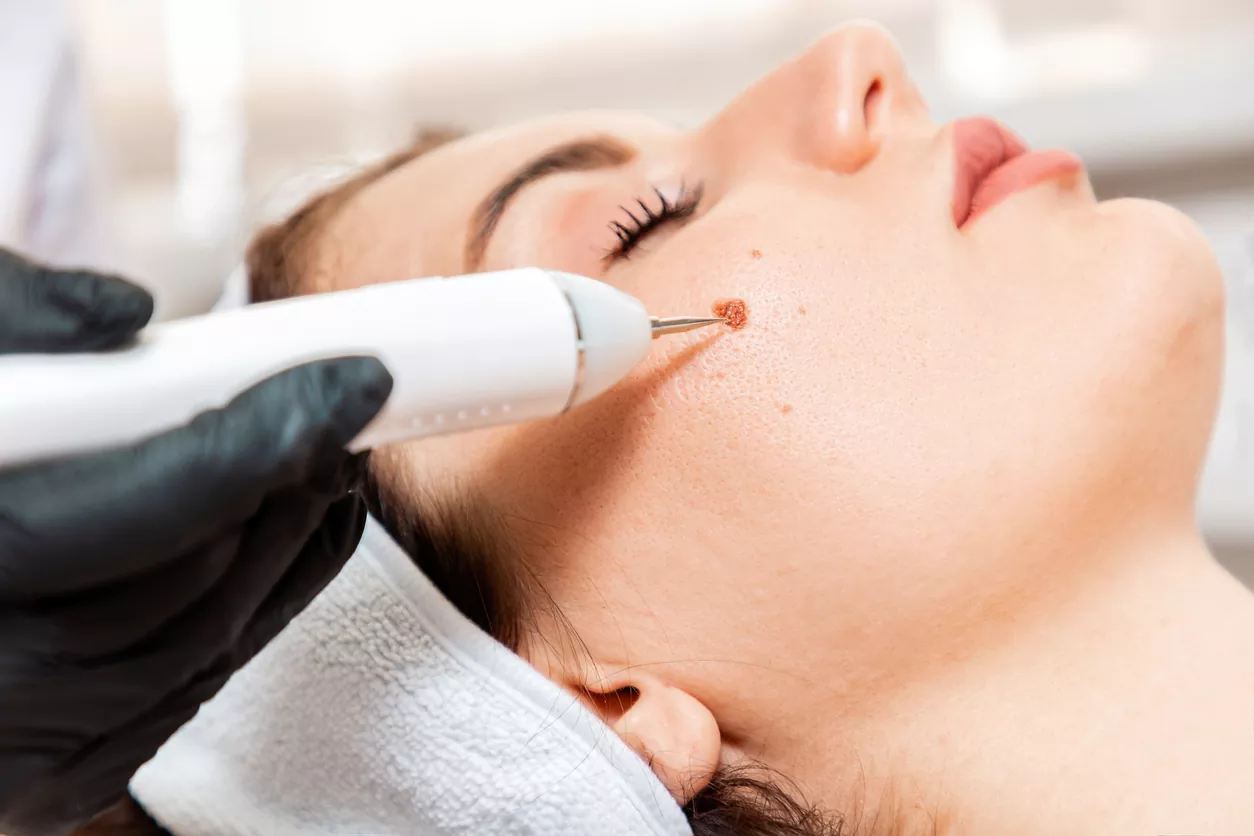
Removal Science: The Modern Clinical Protocol
When a patient chooses to have an accessory tragus removed, the procedure is a study in controlled tissue management. The science of the removal focuses on three main goals: complete excision of the cartilaginous root, aesthetic closure, and minimal tissue trauma.
1. The Geometry of the Incision
Practitioners do not simply "cut off" the tag. Instead, they utilize an elliptical incision. This shape is scientifically chosen because it allows the two edges of the skin to be brought together with minimal tension. By orienting the long axis of the ellipse along "Langer’s Lines" (the natural lines of skin tension in the face), the resulting scar is far more likely to remain flat and eventually become invisible.
2. Management of the Cartilaginous Root
If the cartilage is left behind, it can act as a foreign body, potentially causing a persistent bump or even leading to a minor inflammatory response. The surgical science requires following the cartilage to its terminal point. Once the stalk is isolated, it is truncated at a level below the skin surface so that as the wound heals, the skin "drapes" smoothly over the area without any underlying protrusion.
3. Layered Suture Science
Modern closure often involves two sets of sutures. Deep, absorbable sutures are placed in the subcutaneous layer to "pull" the wound together from the inside. This is a crucial step for Riyadh patients who lead active lifestyles; the internal stitches take the stress off the surface skin, which prevents the final scar from widening over time due to facial movements.
Post-Procedural Healing: The Biological Response
Once the tissue is removed, the body enters a highly programmed healing phase.
-
The Inflammatory Phase (Days 1–3): The body sends white blood cells to the site to clear any debris. Minor redness and swelling are signs of this protective response.
-
The Proliferative Phase (Days 4–21): Fibroblasts begin creating new collagen to bridge the gap. In this phase, the incision may look like a thin pink line.
-
The Remodeling Phase (Months 3–12): The collagen is reorganized and strengthened. In the Riyadh environment, maintaining strict sun protection during this phase is scientifically vital. UV radiation can cause "hyper-melanosis" (darkening) of the fresh collagen, whereas proper care allows the scar to mature into a pale, flat, and nearly imperceptible mark.
Conclusion
The accessory tragus is a small biological footnote in the story of human development, but its management requires a deep understanding of facial anatomy and surgical science. For patients in Riyadh, addressing these tags is not merely a matter of vanity, but an exercise in precision medicine. By utilizing modern techniques that respect the complex embryology and delicate anatomy of the preauricular region, practitioners can provide a permanent, safe, and aesthetically superior result. Whether for a newborn child or an adult seeking to refine their profile, understanding the science of the accessory tragus ensures that the decision to remove it is grounded in medical excellence and a clear expectation of a successful, lasting outcome.